|
moon phases |
Faithfully reality-based
(Fair and Balanced? - nah)
Here follow a number of documents I thought it was worth posting on the web for one reason or another.
- Andrew W Robertson
(Fair and Balanced? - nah)
Here follow a number of documents I thought it was worth posting on the web for one reason or another.
- Andrew W Robertson
Hubblog:
Sketches by Mez
Systema Naturae 2000
In a Small Dark Room
Chris' Memorial Site
Another Dark Little Corner
LJ (mirror)
My Profile
Unashamed Cupidity Roolz
Mail
mail (at) mcpye DOT com

Identity Redacted @ Gleet
ABC (Australia)
 Wondering Minstrels
Windows of the Soul
Tlaloc
New Webdiary
The Bleeder All Year
Gutenberg Project
Scholarpedia
Rainforest site
Hunger site
Make Poverty History
Mr Harden's Artchive
Orwell links
xkcd (comic)
Fave Film Reviews
Flickrzen pictures
Daily Imagery
Unnatural Opera
OzBlogs
Andy W Robertson
Galaxy Zoo (DIY)
Panda's Thumb
Pharyngula
Tangled Bank
Top 10 Global Warming Myths
How to Talk to a Climate Skeptic
SETIS: Australian Texts
SETIS Images
Gnomon Publishing
Recollection Books
Avalon Project, Yale
Idiocentrism
Unclaimed Money
Unclaimed History
 CRN on Wampum/Koufax
RECOMMENDED BLOGS
CRN on Wampum/Koufax
RECOMMENDED BLOGS
Creek Running North
Body & Soul
Nothing New (Bellatrys)
Digby (Hullabaloo)
Making Light
Neil Gaiman's Journal
M Klishis (Random)
Road to Surfdom
Respectful of Otters
Orcinus
Respectful Insolence
Obsidian Wings
Torture Issue
Mbaye Diagne: respect

ARCHIVES
<- To Front Page
October 2002
November 2002
December 2002
January 2003
February 2003
March 2003
April 2003
May 2003
June 2003
July 2003
August 2003
September 2003
October 2003
November 2003
December 2003
January 2004
February 2004
March 2004
April 2004
May 2004
June 2004
July 2004
August 2004
September 2004
October 2004
November 2004
December 2004
January 2005
February 2005
March 2005
April 2005
May 2005
June 2005
July 2005
August 2005
September 2005
October 2005
November 2005
December 2005
January 2006
February 2006
March 2006
April 2006
May 2006
June 2006
July 2006
August 2006
September 2006
October 2006
November 2006
December 2006
January 2007
February 2007
March 2007
April 2007
May 2007
June 2007
July 2007
August 2007
September 2007
October 2007
November 2007
December 2007
January 2008
February 2008
March 2008
April 2008
May 2008
June 2008
July 2008
September 2008
October 2008
November 2008
December 2008
January 2009
February 2009
March 2009
April 2009
May 2009
June 2009
July 2009
August 2009
September 2009
October 2009
November 2009
December 2010
Site Feed (Atom) I used to have a guestbook link here, but it's been swamped with 'comment spam', so regretfully I'm deleting it. You can respond through the comments, I suppose.
mail (at) mcpye DOT com
Sketches by Mez
Systema Naturae 2000
SOME FAVOURITE POSTS
Whitman's Spider
Ratbag on Bronowski
Carl Sagan
Working poor
Paul Eluard (1)
Paul Eluard (2)
War Prayer: 1, 2, 3 Environmental Stories
Shiny! (A cartoon);
A Patient Story;
When Rage Builds;
Big Day
RELATED SITESWhitman's Spider
Ratbag on Bronowski
Carl Sagan
Working poor
Paul Eluard (1)
Paul Eluard (2)
War Prayer: 1, 2, 3 Environmental Stories
Shiny! (A cartoon);
A Patient Story;
When Rage Builds;
Big Day
In a Small Dark Room
Chris' Memorial Site
Another Dark Little Corner
LJ (mirror)
My Profile
Unashamed Cupidity Roolz
mail (at) mcpye DOT com
![]()
![]()
Identity Redacted @ Gleet
ABC (Australia)
Wondering MinstrelsWindows of the Soul
Tlaloc
New Webdiary
The Bleeder All Year
Gutenberg Project
Scholarpedia
Rainforest site
Hunger site
Make Poverty History
Mr Harden's Artchive
Orwell links
xkcd (comic)
Fave Film Reviews
Flickrzen pictures
Daily Imagery
Unnatural Opera
OzBlogs
Andy W Robertson
Galaxy Zoo (DIY)
Panda's Thumb
Pharyngula
Tangled Bank
Top 10 Global Warming Myths
How to Talk to a Climate Skeptic
SETIS: Australian Texts
SETIS Images
Gnomon Publishing
Recollection Books
Avalon Project, Yale
Idiocentrism
Unclaimed Money
Unclaimed History
CRN on Wampum/Koufax
RECOMMENDED BLOGS Creek Running North
Body & Soul
Nothing New (Bellatrys)
Digby (Hullabaloo)
Making Light
Neil Gaiman's Journal
M Klishis (Random)
Road to Surfdom
Respectful of Otters
Orcinus
Respectful Insolence
Obsidian Wings
Torture Issue
Mbaye Diagne: respect

TinyURL: a useful utility
ARCHIVES
<- To Front Page
October 2002
November 2002
December 2002
January 2003
February 2003
March 2003
April 2003
May 2003
June 2003
July 2003
August 2003
September 2003
October 2003
November 2003
December 2003
January 2004
February 2004
March 2004
April 2004
May 2004
June 2004
July 2004
August 2004
September 2004
October 2004
November 2004
December 2004
January 2005
February 2005
March 2005
April 2005
May 2005
June 2005
July 2005
August 2005
September 2005
October 2005
November 2005
December 2005
January 2006
February 2006
March 2006
April 2006
May 2006
June 2006
July 2006
August 2006
September 2006
October 2006
November 2006
December 2006
January 2007
February 2007
March 2007
April 2007
May 2007
June 2007
July 2007
August 2007
September 2007
October 2007
November 2007
December 2007
January 2008
February 2008
March 2008
April 2008
May 2008
June 2008
July 2008
September 2008
October 2008
November 2008
December 2008
January 2009
February 2009
March 2009
April 2009
May 2009
June 2009
July 2009
August 2009
September 2009
October 2009
November 2009
December 2010
Site Feed (Atom) I used to have a guestbook link here, but it's been swamped with 'comment spam', so regretfully I'm deleting it. You can respond through the comments, I suppose.
Article links: To link to a specific posted entry on this blog, click on the hyperlink labelled "[link]" at the end of the article, then use that URL as your link.
Or, if your browser allows, right-click and select "Copy link location", then paste that as your link.
mail (at) mcpye DOT com
Hello Cruel World
Thursday, April 27, 2006
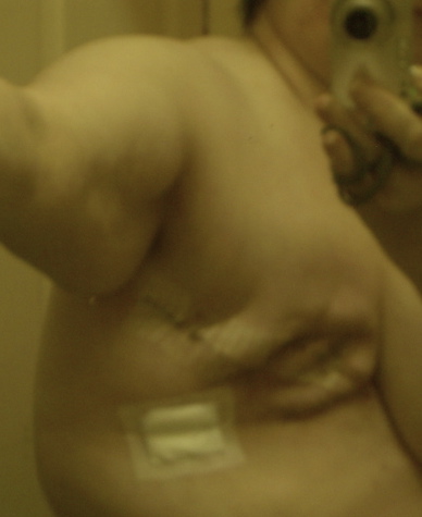
Pathology (Warning: Skip pic if you're sensitive)
Anatomical Pathology — Surgical Pathology Report
Collected April 2006, 12:27Clinical Data
L breast cancer
Nature of Specimen
L modified radical mastectomy
Macroscopic
Labelled "L modified radical mastectomy", the specimen consists of the left breast, 210mm from medial to lateral, 130mm from superior to inferior and 70mm from superficial to deep bearing an ellipse of skin 200x75-4mm with an unremarkable nipple. There is an attached axillary dissection 90x60x20mm. The superficial superior margin is inked blue, the superficial inferior margin green and the deep margin black. The specimen is bi-valved, hinged medially, into superficial and deep halves to reveal a firm irregular grey-white scirrous lesion 45x30x32mm in the 2 o'clock position, 50mm from the nipple in the upper outer quadrant.
The tumour extends to within 18mm of the deep margin (inked black), 8mm of the superficial superior margin (inked blue) in the superficial half and is well clear of the superficial inferior margin. It lies 8mm below the skin. In the deep half of the breast 25mm inferior and deep to the main tumour a second separate firm poorly defined grey tumour 23x15x15mm, is identified on parasaggital sectioning (lesion B), abutting the deep margin and extending to within 30mm of the superficial inferior margin.
Three lymph nodes are involved by tumour, including the most lateral node in the axillary dissection.
... [detailed list of samples & sections ("Blocks") 1A to 1AS ] ... Please see accompanying photograph.
Microscopic
Sections show inflitrating ductal carcinoma (Grade 3). 48x33mm with perineural (Block 1J) and vascular (Block 1I, 1R) invasion. The carcinoma invades to 7mm of the dermis. The skin and nipple are not involved. the second tumour deposit invades skeletal muscle on the deep surface of the specimen and reaches to 3mm from the soft tissue margin
(Blocks 1K, 1M, 1N) and shows vascular invasion. The carcinoma is diffusely strongly positive in the oestrogen receptor positive, and in the cytokeratin 7, focally weakly positive in the progesterone receptor and equivocal (2+) in the HER2. FISH for HER2 has been ordered.
There is focal high grade intraduct carcinoma (Block 1Y).
Metastatic carcinoma is seen in three of eighteen lymph nodes.
Conclusion:
Infiltrating ductal carcinoma (Grade 3): 48x33mm, vascular and perineural invasion, outlying invading skeletal muscle; metastases to three of eighteen lymph nodes.
Left mastectomy and axillary clearance.Addenum
Clinical Details
Breast ca. with modal metastases. HER-2 IHC 2+. HER-2 FISH testing at request of Dr A Field.
Results
HER-2 FISH result (PathVysion HER-2 DNA probe kit): NEGATIVE (Non amplified)
Mean HER-2 copy number per cell: 2.15
HER-2/chr17 ratio: 1.02
(3925) T-04000 M-85003 M-85002
Comments:
Post a Comment

![]()
This is my blogchalk:
Australia, New South Wales, Sydney, English, photography, reading, natural history, land use, town planning, sustainability.